Free Printable Heart Diagram To Label: A Comprehensive Guide for Understanding the Heart’s Anatomy and Function
The heart, a vital organ responsible for pumping blood throughout the body, holds immense significance in human health. Understanding its intricate structure and function is crucial for medical professionals, students, and individuals seeking to maintain cardiovascular well-being. A free printable heart diagram to label serves as an invaluable tool, providing a detailed visual representation of the heart’s anatomy, aiding in the comprehension of its physiological processes.
This guide will delve into the benefits and applications of a printable heart diagram, exploring its role in education, medical practice, and patient understanding. We will provide a comprehensive overview of the heart’s structure, function, and common conditions, equipping readers with a thorough understanding of this essential organ.
Introduction

Alright bruv, listen up! This free printable heart diagram to label is a sick tool for getting your head around the human ticker. It’s like a roadmap for your heart, showing you all the important bits and pieces.
Whether you’re a student trying to ace your biology exam or a healthcare pro brushing up on your anatomy, this diagram is the bomb. You can use it to learn about the heart’s structure, function, and how it keeps you ticking.
Educational Uses
For you students out there, this diagram is a game-changer. It’s the perfect way to visualize the heart and understand how it works. You can use it to study for tests, create projects, or just get a better grasp of human biology.
- Learn the different parts of the heart, like the chambers, valves, and blood vessels.
- Understand how blood flows through the heart and body.
- See how the heart’s electrical system controls its rhythm.
Medical Uses
For healthcare pros, this diagram is a valuable tool for patient education and medical procedures. You can use it to:
- Explain heart conditions to patients in a clear and concise way.
- Help patients understand their treatment options.
- Guide medical procedures, such as cardiac catheterization or heart surgery.
Anatomy of the Heart
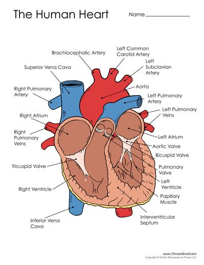
Yo, check it, the heart’s like the OG pump in your body, blud. It’s a muscular organ that’s about the size of your fist, and it’s got this mad system of chambers, valves, and blood vessels that keep your blood flowing like a boss.
Let’s break it down, init. The heart’s got four chambers: two atria (that’s Latin for “rooms”) on top, and two ventricles (also Latin for “rooms”) on the bottom. The atria are like the chill-out zones where blood comes in, and the ventricles are like the powerhouses that pump blood out to your body.
In between the atria and ventricles, there are these valves that act like one-way doors. They make sure that blood flows in the right direction, like a traffic cop for your blood flow.
Now, let’s talk about the blood vessels. The heart’s got two main arteries, the pulmonary artery and the aorta, which are like the motorways of your circulatory system. The pulmonary artery takes blood to your lungs to get some fresh O2, and the aorta takes the oxygenated blood out to the rest of your body.
Major Blood Vessels
- Pulmonary artery: Takes blood to the lungs
- Aorta: Takes oxygenated blood to the body
Valves
- Mitral valve: Between the left atrium and left ventricle
- Tricuspid valve: Between the right atrium and right ventricle
- Aortic valve: Between the left ventricle and aorta
- Pulmonary valve: Between the right ventricle and pulmonary artery
Heart Function
The heart’s primary role is to pump oxygenated blood throughout the body and remove deoxygenated blood from the body. This pumping action is achieved through a coordinated sequence of events known as the cardiac cycle.
The cardiac cycle consists of two main phases: systole and diastole. During systole, the heart contracts and pumps blood out of the heart. During diastole, the heart relaxes and fills with blood.
The cardiac cycle is controlled by electrical impulses that originate in the sinoatrial (SA) node, located in the right atrium. The SA node generates electrical impulses that travel through the heart, causing the atria to contract and fill the ventricles with blood. The electrical impulses then travel to the atrioventricular (AV) node, located between the atria and ventricles. The AV node delays the electrical impulses slightly, allowing the atria to completely fill with blood before the ventricles contract.
The delayed electrical impulses then travel down the bundle of His, a group of fibers that connect the AV node to the ventricles. The bundle of His divides into the left and right bundle branches, which carry the electrical impulses to the left and right ventricles. The electrical impulses cause the ventricles to contract, pumping blood out of the heart.
The cardiac cycle is a continuous process that repeats itself over and over again. The heart rate, which is the number of times the heart beats per minute, is controlled by the autonomic nervous system. The autonomic nervous system can increase or decrease the heart rate in response to the body’s needs.
Cardiac Cycle Flowchart
The following flowchart illustrates the sequence of events in the cardiac cycle:
- The SA node generates an electrical impulse.
- The electrical impulse travels through the atria, causing them to contract.
- The electrical impulse travels to the AV node.
- The AV node delays the electrical impulse slightly.
- The electrical impulse travels down the bundle of His.
- The bundle of His divides into the left and right bundle branches.
- The electrical impulse travels to the left and right ventricles.
- The ventricles contract, pumping blood out of the heart.
- The atria relax and fill with blood.
- The ventricles relax and fill with blood.
- The cardiac cycle repeats.
Common Heart Conditions

Heart conditions are common and can affect people of all ages. Some of the most common heart conditions include coronary artery disease, heart failure, and arrhythmias.
Coronary artery disease is the most common type of heart disease. It occurs when the arteries that supply blood to the heart become narrowed or blocked. This can lead to a heart attack. Symptoms of coronary artery disease can include chest pain, shortness of breath, and fatigue. Treatment options for coronary artery disease include lifestyle changes, medication, and surgery.
Heart failure occurs when the heart is unable to pump enough blood to meet the body’s needs. This can lead to a number of symptoms, including shortness of breath, fatigue, and swelling in the legs. Treatment options for heart failure include lifestyle changes, medication, and surgery.
Arrhythmias are heart rhythm disorders. They can cause the heart to beat too fast, too slow, or irregularly. Symptoms of arrhythmias can include palpitations, chest pain, and dizziness. Treatment options for arrhythmias include medication, surgery, and lifestyle changes.
Applications of a Printable Heart Diagram

A printable heart diagram offers versatile applications across various settings, serving as a valuable resource for students, healthcare professionals, and patients alike.
For students, it provides a tangible and interactive learning tool to grasp the intricate anatomy and functions of the heart. It allows them to visualize and label different heart structures, enhancing their understanding and retention of complex concepts.
For Healthcare Professionals
- Patient Education: Printable heart diagrams can aid healthcare professionals in explaining heart conditions and treatment options to patients in a clear and accessible manner.
- Surgical Planning: Surgeons can use heart diagrams to plan and simulate surgical procedures, ensuring precision and minimizing risks.
- Research and Development: Scientists and researchers can utilize heart diagrams to visualize and analyze cardiac structures and functions, facilitating advancements in medical knowledge and treatment.
For Patients
- Understanding Their Condition: Patients can refer to heart diagrams to gain a better understanding of their heart condition, its symptoms, and treatment options.
- Empowerment: By visualizing their own heart, patients can feel more empowered and engaged in their healthcare decisions.
- Monitoring Progress: Over time, patients can track their progress and recovery by comparing changes in their heart diagram with the help of their healthcare providers.
Resources for Obtaining a Printable Heart Diagram

If you’re in need of a printable heart diagram for educational or research purposes, there are plenty of reputable websites and organizations that offer free downloads.
These resources provide accurate and reliable information, ensuring you have the most up-to-date and scientifically sound information.
Online Resources
- American Heart Association: The American Heart Association provides a comprehensive range of heart-related information, including printable diagrams of the heart’s anatomy and function.
- Mayo Clinic: Mayo Clinic offers detailed and interactive diagrams of the heart, allowing you to explore its structures and functions in an engaging way.
- WebMD: WebMD provides a clear and concise diagram of the heart, highlighting its major structures and blood flow patterns.
Educational Institutions
- Khan Academy: Khan Academy offers free online courses and resources, including a comprehensive lesson on the structure of the heart with downloadable diagrams.
- McGill University: McGill University’s Anatomy Tutorial provides detailed anatomical diagrams of the heart, including cross-sections and three-dimensional models.
- InnerBody: InnerBody offers interactive and visually appealing diagrams of the heart, allowing you to explore its structures and functions in a dynamic way.
Answers to Common Questions
What are the benefits of using a free printable heart diagram to label?
Printable heart diagrams offer numerous benefits, including enhanced comprehension of the heart’s structure, improved visualization of its components, and facilitation of labeling practice, which aids in memorization and retention of anatomical details.
How can I obtain a free printable heart diagram to label?
Several reputable websites and organizations provide free printable heart diagrams. These resources offer accurate and reliable information, ensuring the diagrams are scientifically sound and suitable for educational or medical purposes.
What are some common applications of a printable heart diagram to label?
Printable heart diagrams find applications in various settings, including classrooms for teaching heart anatomy, medical consultations for patient education, and personal reference for individuals seeking to expand their knowledge of the cardiovascular system.